Advancing brain tumour research with preclinical MRI studies
Research on pre-clinical animal models is an essential step in the testing of new treatments. To assess the growth of brain tumours in a non-invasive manner and monitor the physiological effects of new therapies, the NorLux Neuro-Oncology Laboratory in LIH’s Department of Oncology operates an in vivo imaging platform within its germ-free rodent facility. It contains the first small animal scanner in Luxembourg, a device that arises growing interest within the local research community.
Since 2015, LIH owns a Magnetic Resonance Imaging (MRI) instrument developed by MR Solutions that can provide 2D and 3D anatomical and physiological tomographic images of brain and whole body (see images below). This enables the scientists to study the effect of anti-cancer treatments in real-time in clinically relevant animal models.
In addition, the platform allows to perform Optical Imaging and will soon be extended for multimodal molecular imaging with the Positron Emission Tomography (PET) and possibly Single-Photon Emission Computed Tomography (SPECT) technologies. LIH has established a collaboration with the company MR Solutions to further develop innovative MRI pulse sequences.
Although the primary applications of the imaging platform are currently in the field of oncology research, the MRI instrument is also used by LIH researchers from other domains. The applications can be extended to neuro-degenerative, cardiovascular and infectious disease research areas. Thanks to its wide application range, contacts have also been established with other research units from Luxembourg and the Greater Region, interested in using the device.
Dr Olivier Keunen, who is responsible for the in vivo imaging platform, comments: ‘The balance between ease of use and the ability to carry out cutting edge research fits our requirements perfectly. Even users with little or no MRI experience are able to acquire routine scans within minutes using the simple interface “Preclinical Scan”, while advanced tools allow experienced scientists to develop and customise new protocols and pulse sequences for more advanced research.’
Contact and more information here.
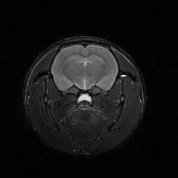
MRI image of a rat brain (ex vivo).
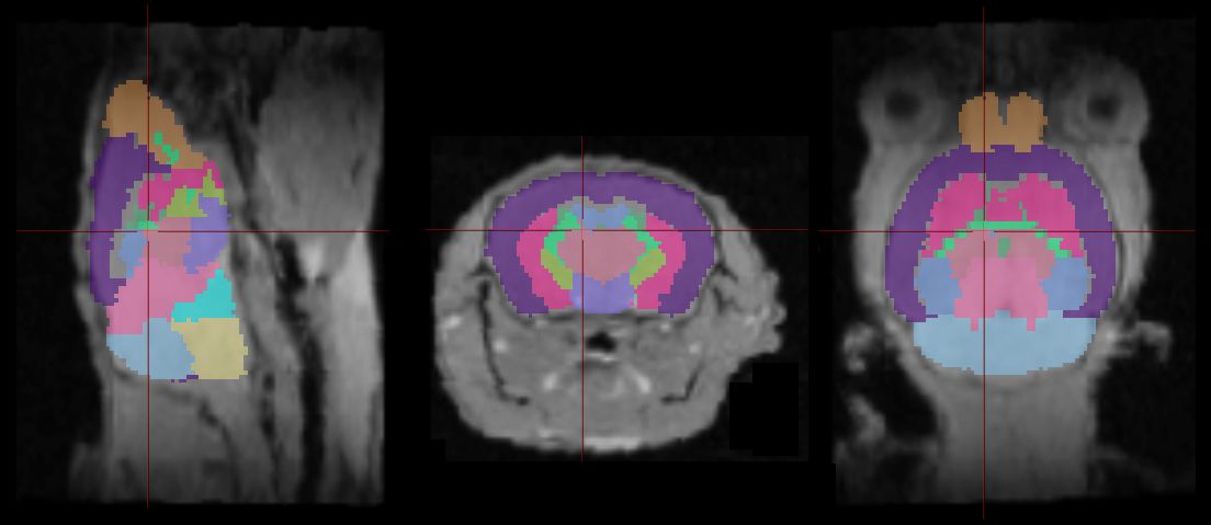
Atlas-based 3D registration and segmentation of a mouse brain